Aesthetics

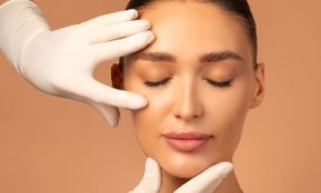
Face
The most modern facial treatments help you look younger, more elegant and happier.

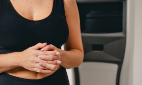
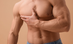
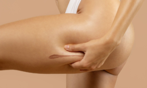
Body
Feel comfortable in your body and correct sources of discomfort.

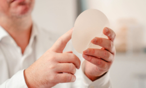
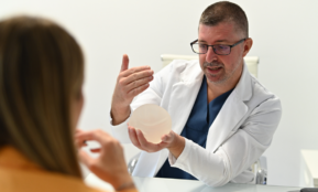
Chest
Make your wish for a better appearance come true and restore your self-confidence with aesthetic correction.

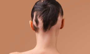
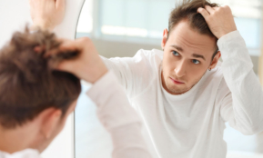
Hair and scalp
Minimally invasive solutions for hair loss and a natural look.

Issues
Find solutions to some of the most common aesthetic problems.
Treatments
VISIA professional facial skin analysis
Dermal fillers
Fractional skin resurfacing
HydraFacial facial cleansing treatment
Chemical peeling
Laser acne treatment
Laser skin rejuvenation
Laser removal of skin redness and capillaries
Laser removal of hyperpigmentation and other pigment changes
Mesotherapy
Micropigmentation of eyelids, eyebrows and lips
Facial rejuvenation with your own blood
Lip augmentation
REVIDERM Skin Peeler microdermabrasion
Skinboosters – restore the skin’s deep glow
Micro needeling tretament
Therapy for facial wrinkles
Vega – bipolar radio frequency
Treatments
Adipology
CoolSculpting cryolipolysis
Dermatological examination
Digital dermatoscopy
Laser hair removal
Removing scars and strech marks
Laser spider vein removal
Pressotherapy
Med Contour body shaping treatment
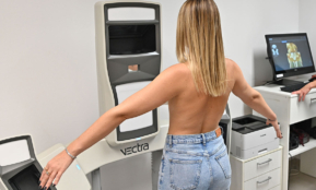
Seca mBCA 515 medical body composition analysis
Treatment for excessive sweating
Venus Legacy multipolar radiofrequency
Z WavePro shock wave therapy
Zerona laser treatment
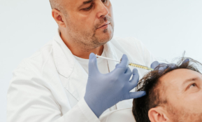
Hair and scalp mesotherapy
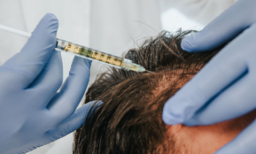
Thinning hair therapy with your own blood

Trichotest – DNA analysis of hair loss and baldness

Hair loss in women – causes and how to prevent it

Hair loss in men – causes and how to prevent it
Alopecia – hair loss
FACE
Acne
Wrinkles
Thin lips
Redness
Deviation of the septum
Spots on the face
Humpback nose
Hyperpigmentation
Pinched ears
Collagen deficiency
Peeling skin
Melasma
Blackheads
Protruding ears
Sagging lower face
Open pores
Scars
Dark circles
Pores
Post-inflammatory hyperpigmentation
Nose reduction
Lowered eyelids
Sunspots
Dry skin
Tired eyelids
Retreating chin
Cleft palate
Big double chin
BODY

Implantology Center
Different methods of solving the problem of missing one or more teeth.

Prosthetics
The most common solution in cases of functional or aesthetic tooth damage.

Aesthetic Dentistry
Harmonious tooth shaping and tooth color correction according to your wishes.

General Dentistry
Modern and timely diagnostics, treatment and dental hygiene are the key to the health of your teeth.
Issues
Find solutions to some of the most common aesthetic problems.

About us

Locations


Almost everyone has moles on their body, which often pose no threat. With regular dermatological check-ups, especially before or after intense sun exposure, there is no reason to remove them.
However, if they start to show signs of change, are in irritable areas, or your dermatologist thinks they should be removed as a preventative measure, this is everything you need to know about mole removal.
Methods of removing moles and benign changes
At the Bagatin Polyclinic, we use the following methods for removing moles and/or benign changes:
- surgical removal
- laser removal of benign changes
- radiofrequency ablation
Surgical and radiofrequency removal methods are available at the Bagatin Polyclinic in Zagreb and Split, while the laser removal method of benign changes is exclusively available in Zagreb.
It should be noted that each removal method has its own advantages and indications/contraindications.
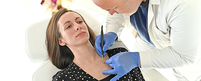
Surgical removal of the lesion is performed under local anesthesia and this method is chosen if a pathohistological analysis of the mole is required, or when we are not completely sure about the benignity of the mole/lesion. After excision, the mole is sent for pathohistological analysis (PHD) in order to establish a diagnosis. When the lesion is removed, the patient has stitches that must be bandaged regularly and are removed after seven to 14 days, depending on the location. After surgical removal, we can expect a scar on the skin, depending on the size of the incision, or the lesion that was removed.

Laser removal is recommended for benign changes, i.e. changes above the skin level. Of course, the patient must first be examined by a dermatologist who then confirms that it is a benign change and that the patient is a candidate for this removal method. Given that laser removal causes tissue destruction, it is not possible to send the mole for PHD analysis. The procedure is also performed under local anesthesia with minimal sensitivity. Unlike surgical removal, the advantage of this method is the precision of the laser and the possibility of minimal invasion of the surrounding tissue, which makes the scar minimal. However, laser removal is only used for benign changes and residual pigment may occur, which is why the procedure must be repeated.
Radiofrequency mole removal is used for benign skin growths. The exceptions are papillomatous moles (benign moles that rise above the skin) and changes above the skin level that can be “flattened” and sent for PHD analysis. Like other methods, this procedure is performed under local anesthesia and is not painful, and it takes about seven days for the wounds to heal (with regular application of antibiotic ointments). After this method of mole removal, the appearance of the scar depends on several factors such as the size of the change, skin reaction and care for the wound itself, so a minimal to moderately large scar can result.
The aforementioned methods can remove all benign skin lesions, including papillomatous and pigmented moles, lipomas, fibromas, hanging warts, small hemangiomas, and keratoses.
During a dermatological examination, a dermatologist will advise you on which method of removing moles and benign lesions is best for you.
Preparation for the procedure
Patients can eat and drink normally before the procedure. It is not recommended to use non-steroidal analgesics four to seven days before the procedure.
Mole excision procedure
The excision site is treated by washing the surgical field with a surgical skin cleanser and then covered with a sterile compress. A local anesthetic is then applied, followed by excision of the lesion. After removal of the desired lesion, surgical sutures are placed and the wound is sterilely bandaged. The client is scheduled for a check-up the next day, and if everything is in order, detailed instructions for home dressing are given.
With laser removal and removal using the radiofrequency method, the desired area is disinfected and a local anesthetic is administered. After the procedure, a dressing is applied for faster recovery.
Frequently asked questions from patients
- Is the procedure painful?
The biggest fear of clients is the pain of the procedure. Before the procedure, local anesthetics are administered to numb the area of interest, minimizing pain during the procedure. Given the type of procedure, patients can expect minimal postoperative pain as the wound’s reaction to the procedure and can take an analgesic if necessary. The administration of anesthesia can also “sting” more than hurt, but this lasts a few seconds during application.
- Is a PHD analysis necessary?
Pathohistological analysis of moles or skin changes is necessary if a dysplastic mole or skin cancer is suspected. The change is sent to the Department of Pathology at KBC, and the results of the analysis are received in two to three weeks. The result can show the type of mole and whether it has been completely removed.
- How visible will the scar be?
After surgical and radiofrequency removal, the scar will always be more visible than with laser removal, but it should be kept in mind that scars fade over time. We recommend that this procedure be performed in the fall and winter months to minimize exposure to sunlight and to prevent post-procedural pigmentation.
-
Am I a candidate for this procedure?
Patients who are not recommended for the procedure are people who react with keloid formation after skin damage, people with autoimmune skin diseases, damaged skin, and skin with inflammation. It is also recommended to avoid radiofrequency mole removal in patients with pacemakers.
Pogledajte naše blogove


